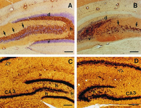
Figure 5.

Photomicrographs of the hippocampus in control (A and C) and x-ray irradiated (B and D) rats. In the irradiated rats, the dorsal blade (d) of the dentate gyrus is greatly reduced, whereas the ventral blade (v) is almost completely missing. Furthermore, the hilar region (h) is smaller, and the Timm-labeled mossy fiber bundle (arrows) is sparse in the irradiated animals. The pyramidal cell layer (p) appears not to be affected either in the regio inferior (CA3) or in the regio superior (open arrow) of Ammon’s horn. Cresyl violet counterstained Timm staining (A and B) and Nissl-like silver staining (C and D). Bar = 200 μm.
