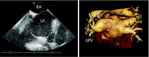
Figure 11 Comparison of MSCT and ICE to evaluate LA and pulmonary vein anatomy. In this patient, both techniques clearly visualise the “common ostium” of the left‐sided pulmonary veins (indicated with the white arrow). Whereas MSCT may provide more detailed three‐dimensional information, ICE also provides online information on the relationship between the ablation catheter and the pulmonary veins during the actual ablation. LIPV, left inferior pulmonary vein; LSPV, left superior pulmonary vein; RA, right atrium.

An official website of the United States government
Here's how you know
Official websites use .gov
A
.gov website belongs to an official
government organization in the United States.
Secure .gov websites use HTTPS
A lock (
) or https:// means you've safely
connected to the .gov website. Share sensitive
information only on official, secure websites.
