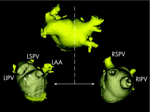
Figure 14 After the fusion process, the “real” anatomy provided by the MSCT scan can be used to guide the actual catheter ablation. With the use of dedicated tools, such as a “clipping plane” (represented by the dotted line), the ostium of the left‐sided pulmonary veins (lower left image) and the right‐sided pulmonary veins (lower right image) can be visualised. The relationship between the ablation catheter and the ostium of the pulmonary veins can be monitored constantly. In this patient, a “common ostium” of the left‐sided pulmonary veins is present. The white arrows on the lower left image indicate the ridge between the left‐sided pulmonary veins and the left atrial appendage (LAA). LIPV, left inferior pulmonary vein; LSPV, left superior pulmonary vein; RIPV, right inferior pulmonary vein; RSPV, right superior pulmonary vein.

An official website of the United States government
Here's how you know
Official websites use .gov
A
.gov website belongs to an official
government organization in the United States.
Secure .gov websites use HTTPS
A lock (
) or https:// means you've safely
connected to the .gov website. Share sensitive
information only on official, secure websites.
