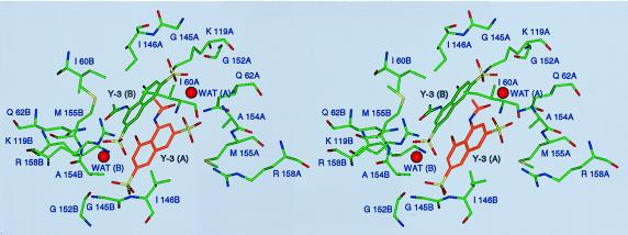
Figure 4.
Stereoview of the Y-3 binding site, shown approximately along the crystallographic twofold axis. Structural elements contributed by either monomer of the enzyme are labeled with A or B, respectively, after the residue number. Water molecules stabilizing the interactions between the two Y-3 molecules, WAT (A) and WAT (B), are shown.

