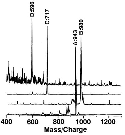
Figure 1.
Laser desorption ionization mass spectra of purified porphyrin intermediates of D. vulgaris cells cultured in the presence of l-methionine-methyl-d3. Peaks A, B, C, and D were assigned to uroporphyrin III octamethyl ester (943), 2,7-methyl-d3 sirohydrochlorin octamethyl ester (980), 2,7-methyl-d3 coproporphyrin III tetramethyl ester (717), and 2,7-methyl-d3 protoporphyrin IX dimethyl ester (596), respectively.

