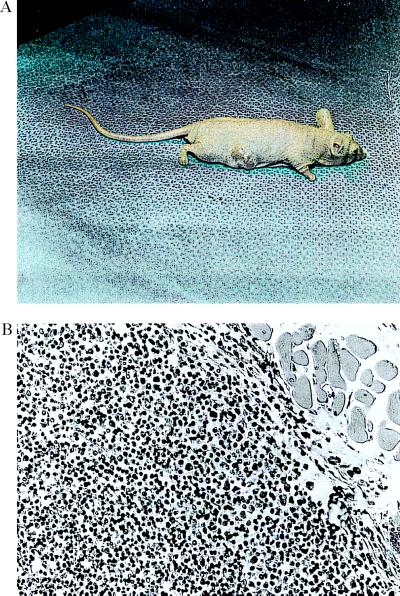
Figure 1.
The data in A and B present a tumor growing in the subscapular area on the back of a nude mouse. This tumor was observed 1 week after receiving a bolus of 5 × 10⁶ cells contained in a 0.1 ml injection into this area of the back. These antisense transfected cells were recovered from four different tumors. (B) Histopathology of a tumor 3 weeks after receiving the cell bolus. The tumor was sectioned into 8-μm sections, fixed in formalin at pH 7. 0, and stained with hematoxylin/eosin. B cell lymphomas containing large lymphoblastoid cells were observed in numerous areas throughout the tumor area. The sections contained a minimal amount of connective tissue. (B, ×200.)

