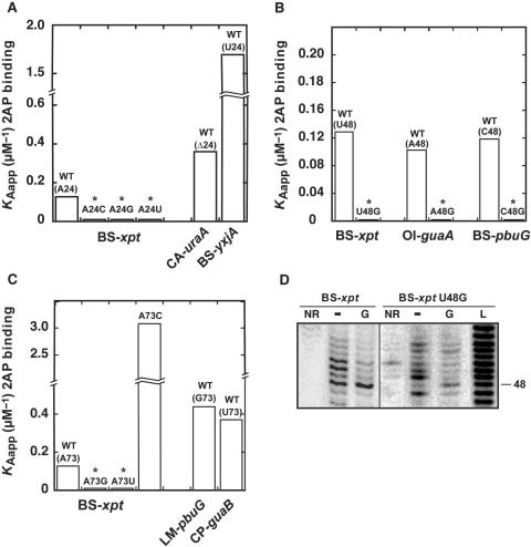
Figure 4.
The importance of nucleotide positions 24, 48 and 73 for 2AP binding. (A) Position 24. 2AP binding affinity histogram for natural variants BS-xpt, CA-uraA and BS-yxjA. Three BS-xpt mutants are also shown where position 24 is changed for a cytosine (A24C), a guanine (A24G) and a uracil (A24U). (B) Position 48. 2AP binding affinity histogram for the three natural variants BS-xpt, OI-guaA and BS-pbuG. Three mutants are also shown in which position 48 is substituted for guanine (BS-xpt U48G, OI-guaA A48G and BS-pbuG C48G). (C) Position 73. 2AP binding affinity histogram for natural variants BS-xpt, LM-pbuG and CP-guaB. Three BS-xpt mutants are also shown where position 73 is changed for a cytosine (A73C), a guanine (A73G) and a uracil (A73U). Asterisks indicate that binding affinity could not be accurately measured as the low 2AP fluorescence quenching due to inefficient binding makes calculation unreliable (KDapp > 25 µM). (D) In-line probing assays of the wild type and the U48G BS-xpt aptamer in absence (−) or in presence of 1 µM guanine (G). Lanes NR and L correspond to molecules that were non-reacted or that were partially digested by alkali, respectively. Position 48 is indicated on the right.

