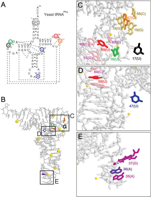
Figure 4.
The s incorporation sites in yeast tRNAPhe and the structure of the tRNA. (A) The secondary structure of the original tRNA transcript. The positions substituted with s are circled. The broken lines show base–base interactions for the 3D structure (37). The boxed G–C pair was changed from the original C–G pair, but this mutation does not significantly alter the original tRNA structure. (B–E) The deep-colored bases were substituted with s, which stacks with the light-colored bases, and the yellow spheres represent Mg2+.

