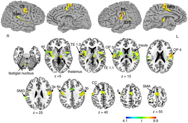
Figure 2. Common activation during voluntary cough, sniff and breathing relative to passive breathing.
Spatially normalized activation was registered and projected onto the single-subject template in the Talairach-Tournoux standard space (group mean activation, p ≤ 0.05, corrected). The color scale illustrates t-values (14 degrees of freedom). CS – central sulcus; SMG – supramarginal gyrus; STG – superior temporal gyrus; SMA – supplementary motor area; OP – operculum; CC – cingulate cortex; T – thalamus; R –right; L - left.

