Figure 3.
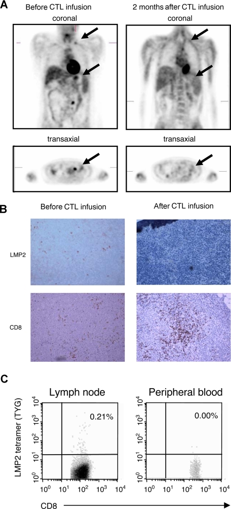
Induction of complete clinical response and LMP2-specific T-cell accumulation at tumor site after CTL infusion. A positron emission tomography (PET) scan demonstrating abnormal fluorodeoxyglucose (FDG) uptake in supraclavicular and para-aortic lymph nodes was observed before CTL infusion in a patient with NHL (pt 2). The follow-up scan 8 weeks after CTL infusion is reported as normal (A). Using immunohistochemistry, CD8+ infiltrating T cells were seen in lymph node biopsy after CTL infusion, which corresponded to a clearance of LMP2+ tumor cells (B). In addition, the percentage CD8+/LMP2 tetramer+ T cells in the lymph node and peripheral blood were compared after CTL infusion by flow cytometry (C). Images were acquired with an Olympus BX41 microscope (OlympusAmerica, Center Valley, PA) with a Plan Achromat 10×/0.25 NA oil objective lens (Olympus, Tokyo, Japan). Cells were stained with hematoxylin (Mayer)-eosin and also with CD8 monoclonal antibody (Dako, Carpinteria, CA) and LMP2 antibody (gift of Dr Friedrich A. Grässer, Institut für Mikrobiologie und Hygiene Abteilung Virologie, Homburg/Saar, Germany) and were used in immunoperoxidase protocol. Images were photographed with an Olympus Q-Color 5 digital color camera using FireWire technology and processed with Adobe Photoshop Elements 2.0 imaging software (Adobe Systems, San Jose, CA). Original magnification, ×10.

