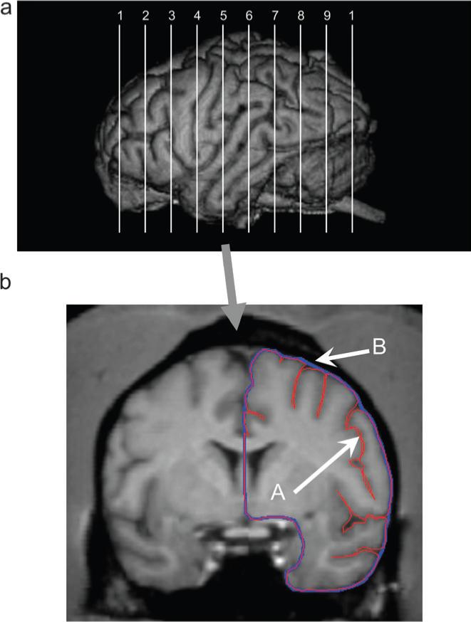
Figure 1.

(a) Upper panel shows the approximate locations in the anterior-to-posterior plane from which the 10 slices and gyrification measures were obtained from each subject. (b) The lower panel shows a single 1-mm slice oriented in the coronal plane on which the contour (A, in red) and outer edge (B, in blue) of one hemisphere are traced using ANALYZE.
