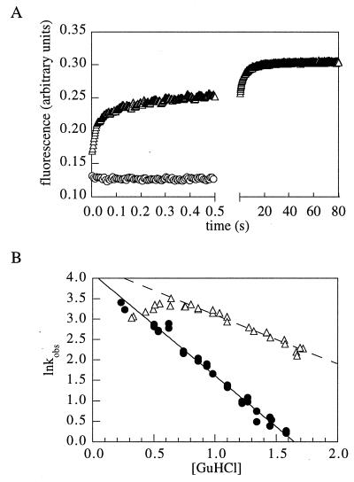
Figure 4.
Anomalous refolding kinetics of variants h-a and h-b. (A) Kinetics of refolding of h-b (triangles, 0.4 M, final GuHCl concentration). The signal of the unfolded protein in 4 M GuHCl is indicated by circles. (B) GuHCl dependence of the logarithm of the folding rate constant for h-a (▵) and wild type (•). The deviation from linearity below 0.7 M GuHCl suggests the accumulation of an intermediate.

