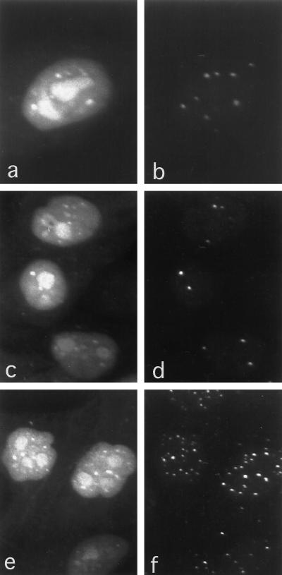
Figure 1.
Confocal microscopy of HEp-2 cells microinjected with FITC–UTP. Fluorescent labeling was localized in PML-containing nuclear bodies and in coiled bodies, both of which are nuclear bodies of unknown function. Although not all PML bodies correspond to FITC–UTP foci, all the foci can be accounted for by either PML bodies or coiled bodies. Note that nucleoli have also incorporated FITC–UTP. Some FITC-labeled foci (a) colocalize with some nuclear bodies labeled with an anti-PML antibody and were detected with an LRSC-conjugated secondary antibody (b). Some FITC–UTP foci (c) colocalize with coiled bodies detected with anti-p80–coilin antibodies and LRSC (d). Cells labeled with both anti-p80–coilin and anti-PML detected by LRSC (f) demonstrate that all of the FITC–UTP foci (e) colocalized with either PML-containing nuclear bodies or coiled bodies (f).

