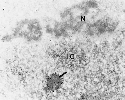
Figure 2.
Ribonucleic acid can be detected in the PML-containing nuclear bodies. Immunoelectron microscopy of a nucleus labeled for PML followed by EDTA-regressive staining (36) demonstrates the localization of RNA in subnuclear structures. Using this technique, a PML body (arrow) demonstrates the characteristic decoration of the nuclear body by PML fringing its outer shell as well as positive staining for RNA throughout the entire nuclear body. As expected, RNA was also detected in nucleoli (N) and interchromatin granule (IG) regions. Chromatin surrounding the nucleolus is devoid of staining. (×20,600.)

