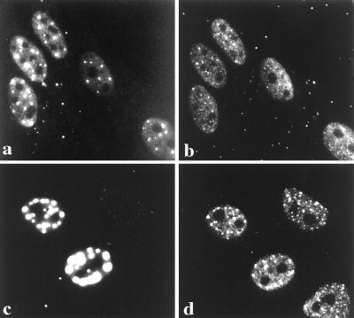
Figure 4.
CBP colocalizes with the PML-containing nuclear body. Double labeling of HEp-2 cells demonstrate PML-containing nuclear bodies as detected with the murine monoclonal antibody 5E10 (a). In the same cells, endogenous CBP is distributed in a finely speckled nucleoplasmic pattern as well as on larger nuclear foci, which can only be observed when utilizing the affinity-purified anti-CBP directed against amino acids 634–648 (b). Comparison of the images demonstrates that CBP and PML colocalize to the same nuclear bodies. To confirm this colocalization, cells exogenously expressing the PML-GFP fusion protein (c) were immunostained for CBP (d), which not only colocalized to the PML-containing nuclear body but appeared to label with a stronger signal, suggesting a recruitment of CBP to the PML bodies.

