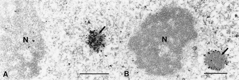
Figure 5.
Ultrastructural analysis of CBP and PML localization in HEp2 cells by immunoelectron microscopy. Silver-enhanced Nanogold pre-embedding immunoelectron microscopy (35) was performed utilizing affinity-purified rabbit antibodies to CBP (aa 634–648) and PML (aa 1–14). (A) CBP was localized in a nuclear body (arrow) with homogeneous labeling throughout. (B) PML localization was restricted to the outer shell of the PML-containing nuclear body (arrow). Nucleoli (N) were unlabeled. (Bar = 0.5 μm.)

