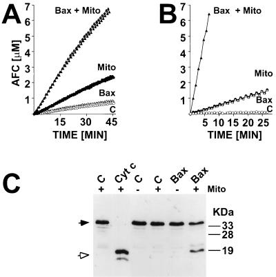
Figure 2.
Bax induces release of mitochondrial factors that trigger processing and activation of cytosolic caspases. In A, supernatants from mitochondria that had been incubated for 1 hr with either 1 μM Bax or diluent control (C) were added to purified cytosol. Alternatively, 1 μM Bax or diluent control were added directly to cytosol. After incubation at 30°C for 0.5 hr, caspase activity was measured by hydrolysis of DEVD-AFC. Data represent micromolar amount of AFC released per time, where samples are normalized for total cytosolic protein content. In B, mitochondria were added to cytosolic extracts with or without 1 μM Bax. After incubation at 30°C for 1 hr, the mitochondria were pelleted by centrifugation and caspase activity was measured in the resulting supernatant. In C, an aliquot of cytosolic fractions was subjected to SDS/PAGE immunoblot assay by using anti-Cyt c antibody. As an additional control (Left), cytosol was treated with 10 μM Cyt c, as described (25). Closed and open arrows indicate the unprocessed zymogen of caspase-3 and the processed form associated with active enzyme, respectively.

