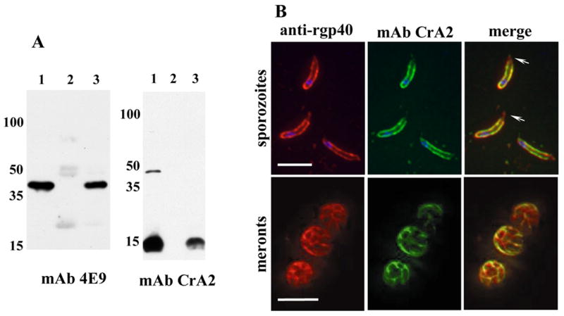
Figure 2. gp40 localizes to the sporozoite membrane by association with gp15.

A. Co-immunoprecipitations: C. parvum GCH1 oocysts were excysted, lysed in low salt lysis buffer (50mM Tris, pH 7.5, 10mM NaCl, 1% TX-100, 1X protease inhibitor cocktail) and proteins precipitated with either rat anti-native gp40 serum or normal rat serum and protein G sepharose beads. After extensive washing, the beads were boiled and the captured proteins analyzed by Western blotting with anti-gp40 mAb 4E9 and anti-gp15 mAb CrA2. Lane 1: total lysate, lane 2: precipitate using normal rat serum, lane 3: precipitate using rat anti-gp40 serum. The high molecular weight band recognized by CrA2 in total lysates is elongation factor EF1α [8, 21]. B. Fixed IFA: C. parvum Iowa isolate sporozoites (top panel) and HCT-8 cells infected with oocysts for 20 hours (meront stage, lower panel) were fixed, reacted with rabbit anti-rgp40 serum and anti-gp15 IgA mAb CrA2, and the primary antibodies co-localized with Alexaflour 594-conjugated goat anti rabbit IgG (Invitrogen, Carlsbad, CA) and FITC-conjugated goat anti mouse IgA (Southern Biotech, Birmingham, AL). Z-stacks with 0.1mm spacing were captured using a Zeiss Axioimager fluorescent microscope and the images deconvolved and merged using Volocity’s iterative restoration program (Improvision, Lexington, MA). Blue fluorescence is nuclei stained with DAPI. Scale bars=5 μm. Arrowheads indicate apical localization of gp40.
