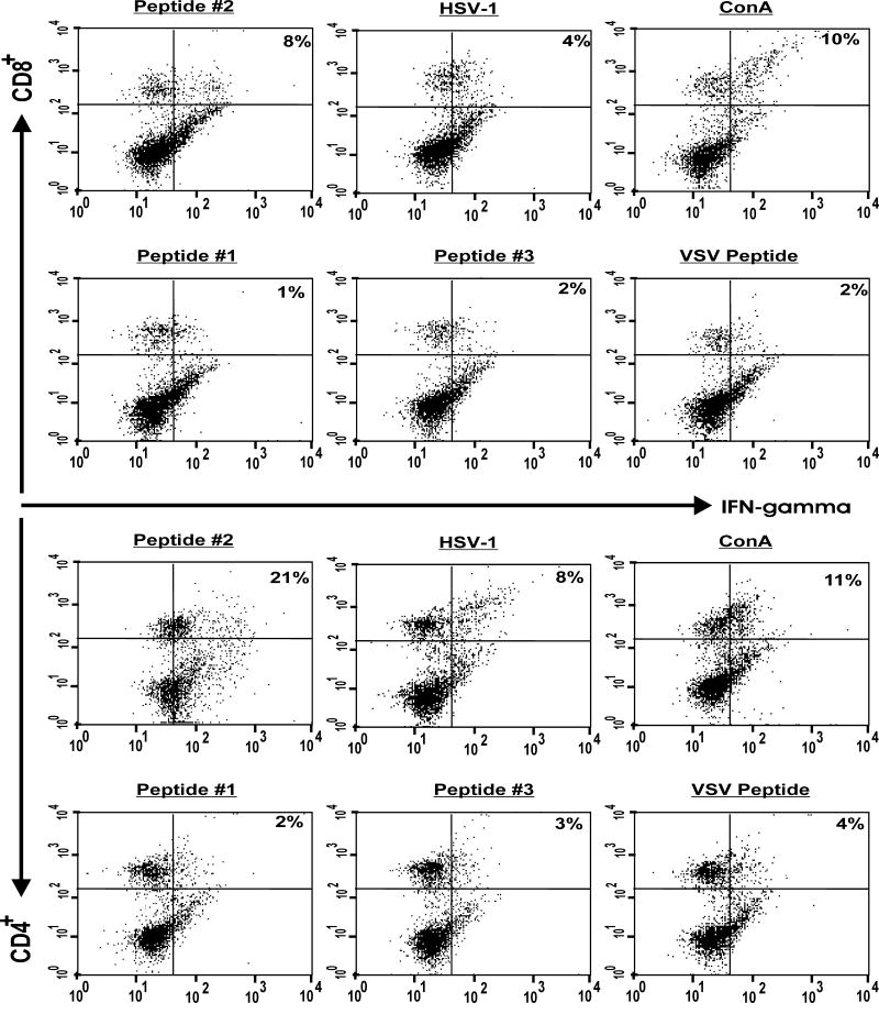
Fig. 2. Generation of CD4+IFN-γ+ and CD8+IFN-γ+ T cells following stimulation of splenocytes from gK-immunized mice with peptide #2.
Mice were immunized with gK, three times as described in Materials and Methods. Three weeks after the third immunization, splenocytes form immunized mice were isolated, and 2 ×10⁶ cells were stimulated with 10 μg of each peptide, 10 PFU/cell of UV-inactivated HSV-1 strain McKrae, 1 μg of ConA, or 10 μg of irrelevant peptide. Four hours before harvesting the cells for FACS analyses, BFA was added as described in Materials and Methods. CD4+ and CD8+ T cells expressing intracellular IFN-γ in response to stimulators were identified using appropriate Abs in a FACSCalibur. The frequency of CD4+ and CD8+ T cells expressing IFN-γ in one representative mouse from gK-immunized mice is shown. Experiments were repeated 2X.

