
Figure 2.
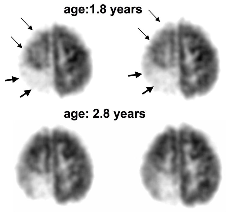
FDG PET scans in girl (patient #6) with a right posterior angioma showing partial recovery after one year. A. The first PET scan at 1.8 years of age showed extensive right hemispheric hypometabolism involving both the right posterior (severe hypometabolism, thick arrows) and superior frontal cortex (mild hypometabolism, thin arrows). B. PET scan one year later showed less extensive hypometabolism with a recovery of frontal cortex glucose metabolism. This improvement in glucose metabolism was accompanied by recovery of hemiparesis in the period between the two scans. Her seizures were relatively well-controlled, with only a single partial seizure during the 1-year period between the two scans.