
Fig. 4.
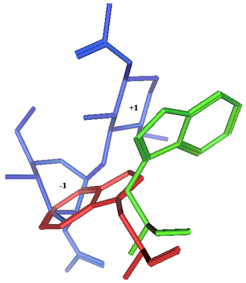
Trp99 of HumGP39 protein along with the binding sugars is shown in stick configuration. Trp99 of HumGP39 from a structure with no sugar bound (PDB code: 1HJX) is shown in red and with sugar bound (PDB code: 1HJW) is shown in green. The bound sugars at −1 and +1 positions are shown in blue. The proteins and sugar are shown in stick configuration