Figure 2. .
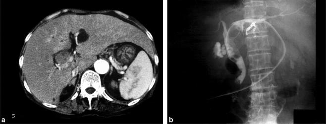
Case no. 2. (a) CT scan shows dilatation of the left hepatic duct and an isodensity mass filling the bile duct. (b) Cholangiography through the PTBD catheter reveals an intraluminal filling defect in the left hepatic duct and common bile duct, which was diagnosed as bile duct cancer at the hepatic hilus.

