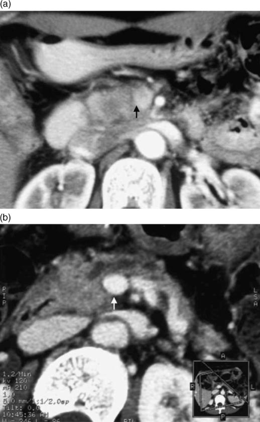
Figure 3. .
A 51-year-old woman with pancreatic head carcinoma. (a) Conventional CT performed at a local hospital revealed a low-density area in the head of the pancreas and the boundary to the portal vein was unclear (arrow). At that hospital, the tumor was diagnosed as inoperable due to portal vein invasion. (b) MPR (multi-planar reconstruction) images obtained by MDCT revealed that the portal vein was intact. Pancreatoduodenectomy combined with resection of the portal vein was performed. Histopathological examination showed no invasion to the portal vein (arrow).

