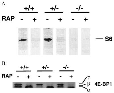
Figure 3.
In vivo phosphorylation of S6 and 4E-BP1. (A) S6 phosphorylation in vivo. Cells were incubated with [32P]orthophosphate in phosphate-free medium in the presence or absence of rapamycin (10 ng/ml) for 3 hr. Cells were lysed in hypotonic buffer, and ribosomes were enriched by ultra centrifugation using a sucrose cushion. Ribosomal proteins were then separated on SDS/10% polyacrylamide gels, and phosphorylated S6 (≈32 kDa) was visualized by using the PhosphorImager and image quant analysis (Molecular Dynamics). (B) In vivo phosphorylation status of 4E-BP1. Cells were treated with vehicle or rapamycin as described in Fig. 1C. Protein extracts were separated on SDS/15% polyacrylamide gels and immunoblotted by using a rabbit polyclonal antibody raised against 4E-BP1. The band with the highest mobility (α) corresponds to hypophosphorylated 4E-BP1, and the bands with lower mobility (β, γ) correspond to hyperphosphorylated 4E-BP1 (23, 24, 28).

