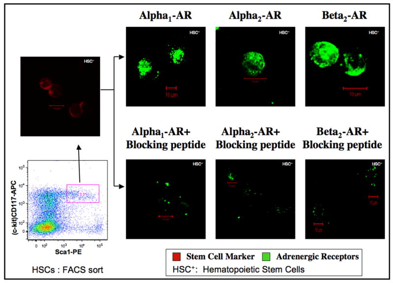
Figure 5.

Adrenergic receptor distribution in murine hematopoietic stem cells (HSCs). Murine bone marrow cells were depleted of lineage-committed cells using Ab coupled micro-bead technology followed by dual staining with anti Sca-1- PE and anti CD117- APC antibodies. Subsequently, CD117high, Sca-1+ population were sorted by flow cytometry to yield HSCs (Lin−, CD117high, Sca-1+) represented in boxed area of the dot plot. 3.31% of Lin− cells are shown (red) in the confocal image right above the boxed area and representing HSCs. Aliquots of HSCs were labeled with primary antibody specific for adrenergic receptor subtypes with and without specific blocking peptides followed by FITC conjugated secondary antibody. Confocal images were obtained in a Zeiss LSM 510 confocal microscope. Green color represents binding of the polyclonal Abs to the C-terminus of adrenergic receptor subtypes. Image resolution=1024 × 1024; Scale bar=10μm.
