
Fig. 3.
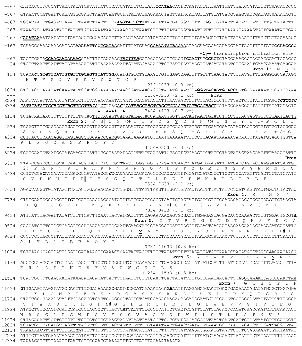
Nucleotide sequence and structural features of M. sexta PAP-1 gene. Nucleotides in the 5′-flanking region are assigned negative numbers. Nucleotide 1 is assigned based on the primer extension results (Fig. 2). Exon sequences are underlined with the encoded amino acid sequences listed below translated exons, using the one-letter code under the 2nd nucleotide of each codon. While some regions of the intron sequences (marked “---”) are not shown, their sizes and positions are indicated. GATA boxes (6-nucleotide) and ISRE sites (13-nucleotide), bold and double underlined; NF-κB motifs (10-nucleotide) and EcRE (15-nucleotide), bold and single underlined; TATA boxes (6-nucleotide) and an imperfect inverted repeat (78 nucleotides), bold italic and double underlined. Mismatches in the repeat are marked with “▲”. Single nucleotide polymorphic sites are in bold italic on the DNA sequence. Among them, nonsynonymous substitutions are further indicated on the affected amino acid residues (bold and underlined). The Cys residues in the clip domain and the catalytic residues in the SP domain are indicated with “+” and “#”, respectively.