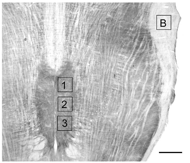
Figure 1.

Photomicrograph of a horizontal section through the medulla stained for 5HT2A immunoreactivity. Rostral (1), middle (2) and caudal (3) sample areas are shown together with the location of the sample area analyzed for background label (B). Scale bar = 750 μm.
