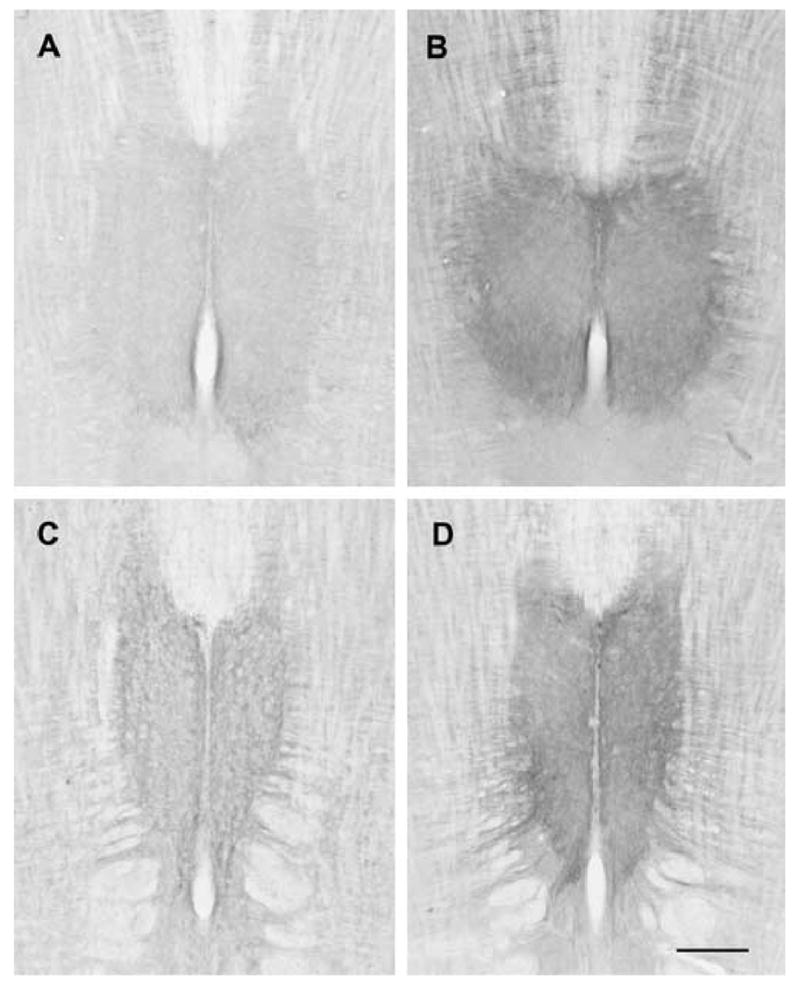
Figure 3.

Photomicrographs of horizontal sections through the hypoglossal nucleus of young and old female rats stained for 5HT2A immunoreactivity. A. Young female, dorsal section. B. Old female, dorsal section. C. Young female, ventral section. D. Old female, ventral section. Sections from young and old rats were reacted at the same time. Images were captured using identical illumination parameters; no further adjustments were made. Scale bar = 750 μm.
