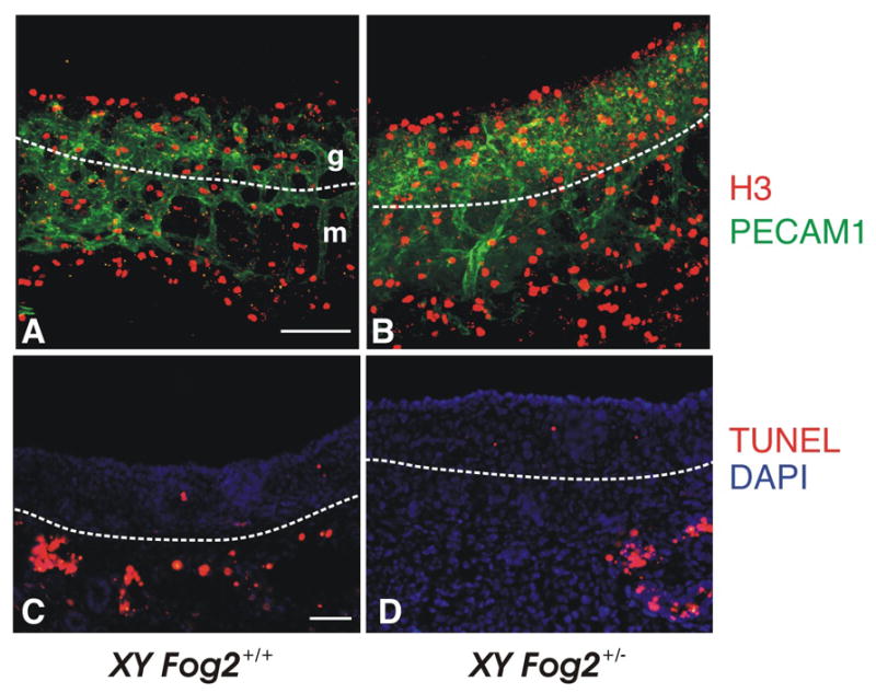
Figure 8.

Comparison of the proliferation and apoptosis in the E11.5 XY gonads isolated from control (A, C) and Fog2 heterozygous (B, D) embryos. The gonads were stained with anti-PECAM1 antibody (green, A-B) and phosphorylated histone H3 (red, A-B). Frozen sections of gonad-mesonephros complex were stained to detect apoptotic cells/bodies (red; nick-end labeling, TUNEL) (C-D). No significant change in the proliferation (A-B) or apoptosis (C-D) are observed in the Fog2+/− samples compared to the control XY gonads. Apoptotic cells are easily detected around mesonephric tubules (arrows in D-F) as has been previously reported (e.g. (Allard et al., 2000; Kim et al., 2006). The white dotted line indicates the boundary between mesonephros (m) and gonad (g). The scale bar is 100 μM (A-B) and 50μm (C-D).
