
Figure 3.
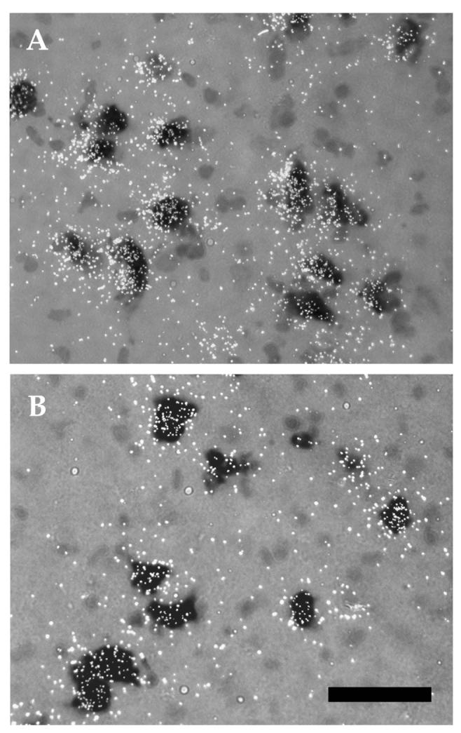
Photomicrographs of α6 mRNA expression within midbrain DA neurons at P21 (A) and adult (B). Note the higher number of silver grains per DA neuron (dark cells) in P21 as compared to the adult. The peak of expression of this subunit at P21 is, therefore, due to higher level of mRNA per DA neuron. Scale bar = 50μm.