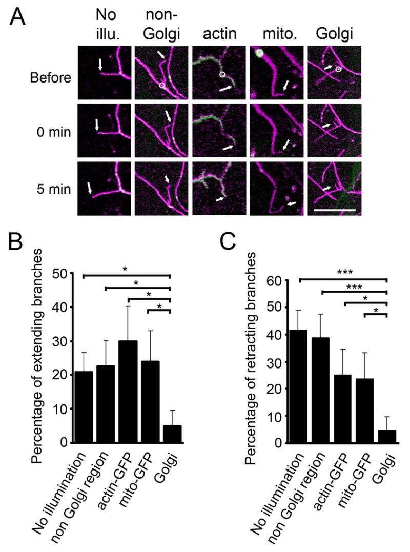
Figure 5.

Laser damaging of dendritic Golgi outposts reduces the extension and retraction of dendritic branches. (A) Examples of dendritic branches without laser illumination (No illu.), after focal intense laser illumination on regions lacking Golgi outposts (non-Golgi), on regions with actin-GFP (actin), on mitochondria (mito.), and on Golgi outposts (Golgi). Magenta: mCD8-dsRed. Green: actin-GFP in “actin”, mito-GFP in “mito.”, GalT-EYFP in the other 3 panels. Upper, middle, and lower panels represent images before, immediately after, and 5 minutes after laser damaging, respectively. Circles indicate the regions of laser illumination. Arrows point to the tips of the branches being tested. Scale bar: 10 μm. (B–C) Percentage of branches that extended (B) or retracted (C) after each manipulation.
