
Fig. 1.
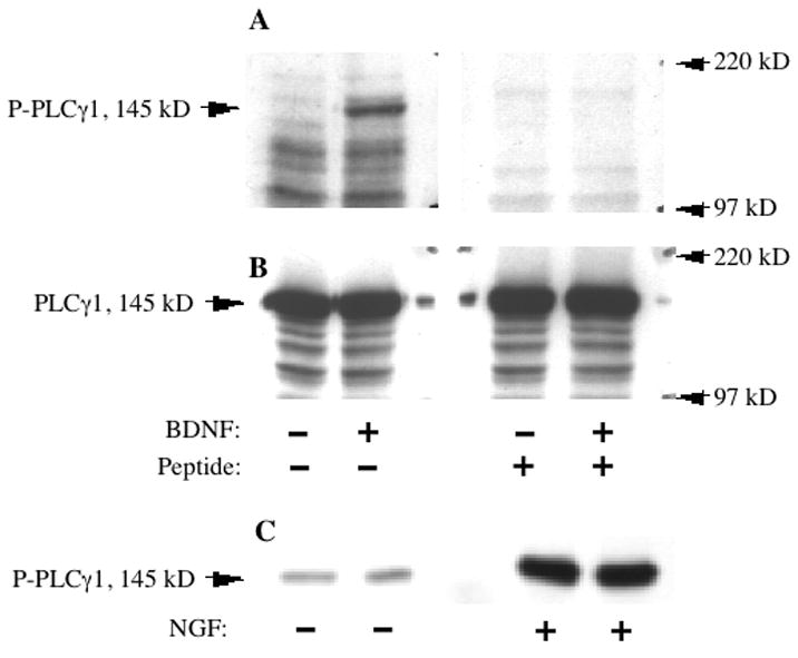
Specific detection of neurotrophin-activated PLCγ1 by a phospho-PLCγ1 antibody. A: Western blots of cell lysate from primary striatal neurons, treated with or without 50 ng/mL BDNF for 10 min. Both blots were probed with the phospho-PLCγ1 antibody (P-PLCγ1), but the antibody used on the right-hand blot was pre-incubated with the antigenic phospho-peptide used to generate it. This pretreatment blocked the detection of BDNF induced PLCγ1 phosphorylation observed in the blot on the left. B: The same membrane as in A, reprobed with an antibody against total PLCγ1, demonstrating the abundant and equal presence of total PLCγ1 in each lane. C: Duplicate lanes of lysate from PC12 cells treated with or without 50 ng/mL NGF for 10 min, and then immunoprecipated with a highly specific mixed monoclonal PLCγ1 antibody. Immunoprecipitated PLCγ1 is detected by the phospho-PLCγ1 antibody.