Figure 3.
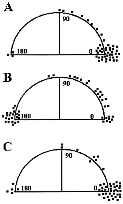
Cell orientational changes during exposure to various solutions in the microscope stopped-flow chamber. (A) Neutrophil orientation changes after pulsed exposure to HBSS in the absence of FMLP. No significant changes are observed. (B) When neutrophils are exposed to a temporally decreasing FMLP signal, most cells reorient at 180° relative to the initial direction of polarization. (C) When neutrophils are exposed to a temporally increasing series of injections, reorientation is not observed. Experiments were replicated on 4–14 separate days.

