Figure 1.
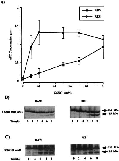
Concentration-dependent activation of caspase 3 activity by GSNO and subsequent time-dependent PARP cleavage in RAW and RES cells. (A) RAW and RES were treated for 8 h with the indicated concentrations of GSNO. Cell extracts (50 μg) were then incubated in the presence of the fluorescent caspase 3 substrate DEVD-AFC (25 μM) for 1 h at 32°C. Caspase 3 activity was measured fluorometrically after substrate cleavage with excitation at 400 nm and emission at 505 nm. Values are the mean ± SD of four experiments. (B and C) PARP cleavage was detected by Western blot analysis using a polyclonal antibody specific for PARP. The arrows indicate the 116-kDa PARP protein and its 85-kDa cleavage product. Time-dependent PARP cleavage after incubation of RAW and RES cells with either 200 μM GSNO (B) or 1 mM GSNO (C) is shown. Results are representative of three experiments.

