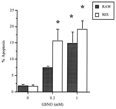
Figure 2.
GSNO-induced apoptosis in RAW and RES cells. Cells were treated for 8 h with 200 μM or 1 mM GSNO, respectively. Apoptosis was quantified by flow cytometry using propidium iodide exclusion. Approximately 10,000 cells were sampled, and the percentage of cells undergoing apoptosis was calculated. Data are representative of results from at least six similar experiments.

