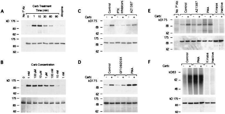
Figure 1.
Muscarinic receptor signaling activates PYK2. (A–D) Cells expressing PYK2-HSV were treated as indicated. PYK2 was immunoprecipitated from cell lysates with anti-HSV antibody, and determined by anti-phosphotyrosine Western blot analysis (Upper). (A) Cells were treated at the indicated timepoints with 50 μM carbachol. Controls omitting the primary antibody (Left) or testing atropine pretreatment (1 min, 5 μM; Right) reflect a 10-min carbachol stimulation. (B) Cells were treated with the indicated concentrations of carbachol for 2 min. (C) Cells were either untreated (−) or treated (+) with 50 μM carbachol for 2 min. Pretreatment with PTK inhibitors was for 20 min at these concentrations: genestein, 10 μM; lavendustin A, 1 μM; tyrphostins A23, A25, and A48, 1 μM each. Pretreatment with 5 μM A21387 was for 15 min. (D) Cells were untreated (−) or treated (+) with 50 μM carbachol for 2 min. Pretreatment with 5 μM GF109203X was for 20 min. Pretreatment with 1 μM PMA was for 15 min. (E and F) Autoradiographic analyses of immune complex kinase assays of PYK2 (Upper). Lysates of cell lines expressing either PYK2-HSV or kinase inactive PYK2-HSV prepared before (−) and after (+) 50 μM carbachol stimulation were immunoprecipitated with anti-HSV antibody and the immune complexes used for in vitro kinase assay in the absence (E) or presence (F) of exogenous poly(Glu,Tyr)4:1 polypeptide substrate. Kinase reactions were stopped with 2× SDS/PAGE sample buffer, the mixtures subjected to SDS-PAGE, and the gels dried for autoradiography. In some cases, the cells were pretreated for 15 min with either 5 μM Ca2+ ionophore A21387 (E) or with 1 μM PMA (E and F). A fraction of each immunoprecipitation was reserved for Western blot analysis with anti-HSV antibody to ascertain the equivalency of PYK2 present in immunoprecipitates (A–F Lower).

