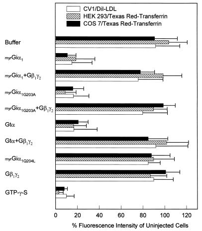
Figure 3.
Endocytosis of fluorescent ligands. Cells were injected with solutions containing 50 μM G protein subunits or 10 mM GTP[γS] as described in Fig. 2, except fluorescein-conjugated dextran was omitted. Two hours after injection, COS-7 cells and HEK-293 cells were incubated with 20 μg/ml FITC-transferrin, and CV1 cells were incubated with 5 μg/ml DiI-LDL for 4 min at 37°C. Quantitation of the internalized fluorescent ligands is described in detail under Materials and Methods.

