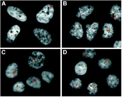
Figure 6.
Dual-label FISH analysis of XIST RNA and X-specific α-satellite DNA. To examine further the disperse localization of XIST RNA in hybrid cells, the XIST signal (red) was detected as in Fig. 5, and the cells then were fixed in paraformaldehyde for subsequent denaturation and detection of X-specific α-satellite sequences (green). DAPI staining of nuclei is represented in gray scale (see Materials and Methods). (A) Normal female fibroblasts. (B) The inactive X human–hamster hybrid X8–6T2S1. (C) The inactive X human–hamster hybrid 8121-TGRD. (D) The XIST reactivant hybrid clone 4F5.

