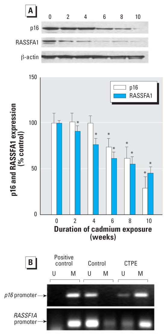
Figure 4.
Expression and promoter region methylation status of p16 and RASSF1A during chronic cadmium exposure. Cells were grown in the presence or absence of 10 μM cadmium for up to 10 weeks. (A) Proteins were isolated and subjected to Western blot analysis using antibodies against p16, RASSF1A, and β-actin. Blots were analyzed densitometrically, normalized to β-actin, and expressed as percent of control. (B) Methylation of the p16 and RASSF1A promoter regions determined by MSP. The presence of visible PCR product in lanes marked “U” indicates the unmethylated genes, whereas the presence of product in lanes marked “M” indicates the methylated gene. The source of DNA is indicated above each lane. CpGENOME universal methylated DNA (CHEMICON International, Inc., Temecula, CA) was used as a positive control.

