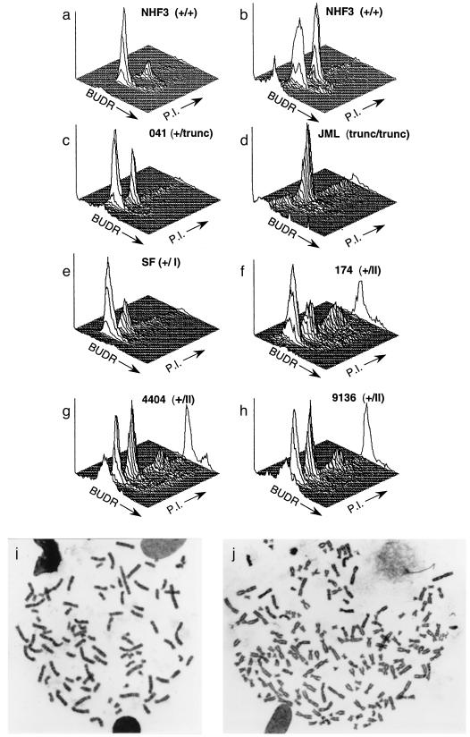
Figure 1.
Analysis of the status of the spindle cell cycle checkpoint in human fibroblasts. Cell cycle was analyzed by flow cytometry to determine the distribution of DNA content in NHF3 incubated without (a) or with (b) 500 ng/ml of colcemid for two population doublings. With increasing time in colcemid, an increasing fraction of normal cells accumulate in the G2/M peak. LFS fibroblasts (c–h) are described in Table 1 and ref. 24. LFS cells were processed for cell-cycle distribution of DNA content as for NHF, except for JML cells, which were incubated for 1 population doubling time. The three-dimensional plots represent cell number (y axis) versus BrdUrd (BUDR) incorporation (z) and PI (x). Plots show 104 cells. (i and j) Metaphase spreads from NHF cells expressing a p53RSC mutation (p53–143A) after two population doubling times in colcemid. Metaphase spreads with 92 (i) and 182 (j) chromosomes are presented.

