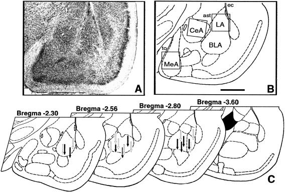
Figure 1.
Cannula placements. (A) Photomicrograph of Nissl-stained section of left amygdala in rat injected with APV. (B) Schematic representation of the same section. Inserted squares represent the areas shown on Figure 8 for examples of c-Fos immunostaining. Scale bar, 1 mm; (to) tractus opticus; (ec) external capsule; (ast) amigdalostriatal area. (C) Cannula tip placements from rats infused with vehicle (open arrows) or 5 μg of APV (filled arrows). Only rats with cannula tips at or within the boundaries of basolateral nuclei were included in the data analysis.

