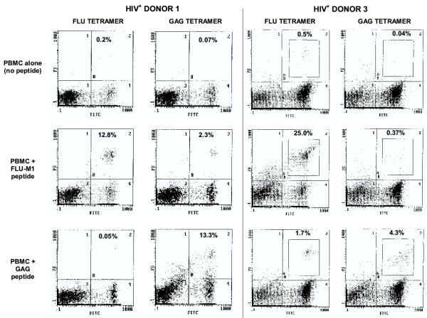
Figure 2.
Tetramer analyses of lymphocytes from HIV-seropositive donors. After in vitro stimulation with IL-2 plus either FLU-M1:58–66 or HIV p17 GAG:77–85 peptide (or no peptide as background), lymphocytes from HLA-A2+, HIV-seropositive Donors 1 and 3 were stained with FITC-labeled anti-CD8 mAb and either HLA-A2/FLU-M1:58–66 tetramer or HLA-A2/GAG:77–85 tetramer (both PE-labeled), as indicated, and viable cells were analyzed by flow cytometry (nonviable cells positive for staining with the dye 7-AAD were excluded). Percentages of CD8+ cells that stain with tetramer appear in the upper-right quadrant of the histograms.

