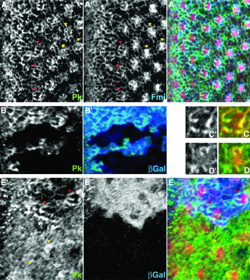
Fig. 5. Pk localization during polarity establishment in eye development. Confocal images of dorsal sides of third instar eye disks stained for Pk (green) are shown. (A), (C) and (D) show a wild-type disk; (B) and (E) show disks with pknull and stbmnull clones, respectively. In (A) and (E), F-actin (stained with phalloidin; red) is shown as reference for the location of ommatidial preclusters. Morphogenetic furrow is at the left margin of each panel, except for (C) and (D). (A) Pk (A′) and Fmi (A”, blue in merged image) colocalize in ommatidial preclusters in R3 and/or R4 cells. Examples in row 5 (red arrowheads) and around row 7 (yellow arrowheads) are highlighted [see also (C) and (D)]. (B) The antibody against Pk is specific, as staining is absent from pk– clones (marked by absence of βGal staining, blue in B′). (C and D) Single photoreceptor clusters of ommatidial row 5 (C) and row 7 (D) show the ‘double-horseshoe’ and the horseshoe-like pattern of Pk [green and (C′), (D′)], respectively. Fmi (red) colocalizes with Pk. (E) stbmnull mutant clones (marked by absence of blue βGal staining in E”); Pk staining is very diffuse. In more mature clusters, partial membrane association can sometimes be detected (compare yellow arrowheads with one another and with red arrowheads).

An official website of the United States government
Here's how you know
Official websites use .gov
A
.gov website belongs to an official
government organization in the United States.
Secure .gov websites use HTTPS
A lock (
) or https:// means you've safely
connected to the .gov website. Share sensitive
information only on official, secure websites.
