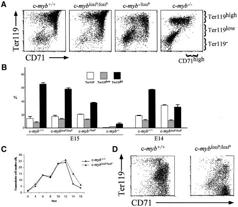
Fig. 4. Erythroid differentiation is perturbed in the presence of reduced levels of c-Myb. (A and B) Flow cytometric analysis of fetal liver cells from wild-type, c-mybloxP/loxP, c-myb–/loxP and c-myb–/– embryos stained with Ter119–biotin and anti-CD71 followed by streptavidin–PE and anti-IgG1–FITC. The profiles in (A) are derived from E15 embryos. The summary histogram in (B) includes data from E15 embryos and also similar stainings of fetal livers from wild-type and c-mybloxP/loxP E14 embryos. (C and D) Culture of wild-type and c-mybloxP/loxP E14 fetal liver cells under conditions favouring expansion of erythroid precursors, maintaining a cell concentration of 1–3 × 10⁶/ml. (C) The cumulative cell number. (D) Cultured cells were stained with Ter119-biotin and anti-CD71 as described in (A) at day 8.

An official website of the United States government
Here's how you know
Official websites use .gov
A
.gov website belongs to an official
government organization in the United States.
Secure .gov websites use HTTPS
A lock (
) or https:// means you've safely
connected to the .gov website. Share sensitive
information only on official, secure websites.
