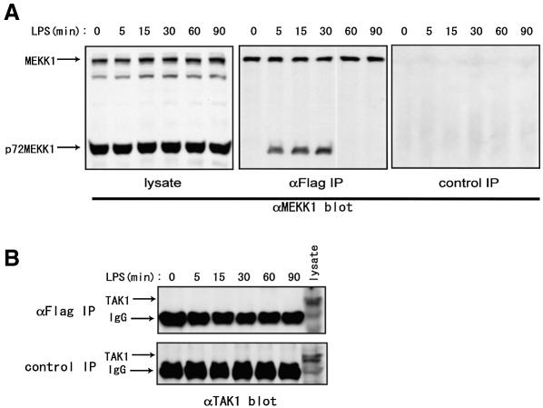
Fig. 6. Complex formation of JIP3 and MEKK1. RAW264.7 cells stably expressing Flag-tagged JIP3 were stimulated with LPS for the indicated times. (A) The anti-MEKK1 immunoblot of the cell lysates is shown in the left panel. For the immunoprecipitation experiments, anti-Flag or control antibody was cross-linked to protein A beads and incubated with the cell lysates. The immunoprecipitates were separated by SDS–PAGE and probed with anti-MEKK1 antibody (two right panels). (B) Anti-Flag and control antibody immunoprecipitates were separated by SDS–PAGE and probed with anti-TAK1 antibody.

An official website of the United States government
Here's how you know
Official websites use .gov
A
.gov website belongs to an official
government organization in the United States.
Secure .gov websites use HTTPS
A lock (
) or https:// means you've safely
connected to the .gov website. Share sensitive
information only on official, secure websites.
