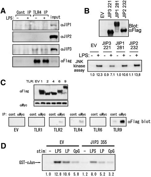
Fig. 7. Involvement of JIP3 in other TLR signals. (A) HEK 293 cells were transiently transfected with p3XFlag-CMV14-TLR4, pcDNA3.1(+)-mCD14, and pcDNA3.1(+)-mMD-2. At 48 h after the transfection, cells were left untreated or stimulated with 1 µg/ml LPS for 20 min and lysed. At 48 h after transfection, cells were lysed and immunoprecipitates of anti-Flag and control antibodies were separated by SDS–PAGE. Immunoblotting was performed with anti-JIP1, anti-JIP2, anti-JIP3 or anti-Flag antibody. (B) HEK 293 cells were transiently transfected with pEEBOS-Flag-JIP3 221, pEFBOS-Flag-JIP1 281 or pEFBOS-Flag-JIP2 232, along with p3XFlag-CMV14-TLR4, pcDNA3.1(+)-mCD14, p3cDNA3.1(+)-mMD-2 and pcDNA3.1(+)-HA-JNK1. At 48 h after the transfection, cells were left untreated or stimulated with 1 µg/ml LPS for 20 min, and lysed. Immunoblotting results using anti-Flag antibody are shown in the upper panel. Anti-HA immunoprecipitates were tested for in vitro kinase assay on GST–cJun5-89 as the substrate. (C) 293T cells were transiently transfected with an expression plasmid of Myc-tagged JIP3 in combination with the indicated expression plasmid of Flag-tagged TLR. At 48 h after transfection, cells were lysed and protein expression was examined using anti-Flag and anti-Myc antibodies (upper two panels). JIP3 was immunoprecipitated with anti-Myc antibody. Coprecipitated TLRs were detected by anti-Flag antibody. (D) RAW264.7 cells stably transfected with the empty vector or the expression plasmid of JIP3 1–355 were treated with 1 µg/ml LPS, 10 μg/ml synthetic lipoprotein, or 1 µM CpG ODN for 20 min and anti-JNK1 antibody immunoprecipitates were examined for their kinase activity on GST–cJun5-89.

An official website of the United States government
Here's how you know
Official websites use .gov
A
.gov website belongs to an official
government organization in the United States.
Secure .gov websites use HTTPS
A lock (
) or https:// means you've safely
connected to the .gov website. Share sensitive
information only on official, secure websites.
