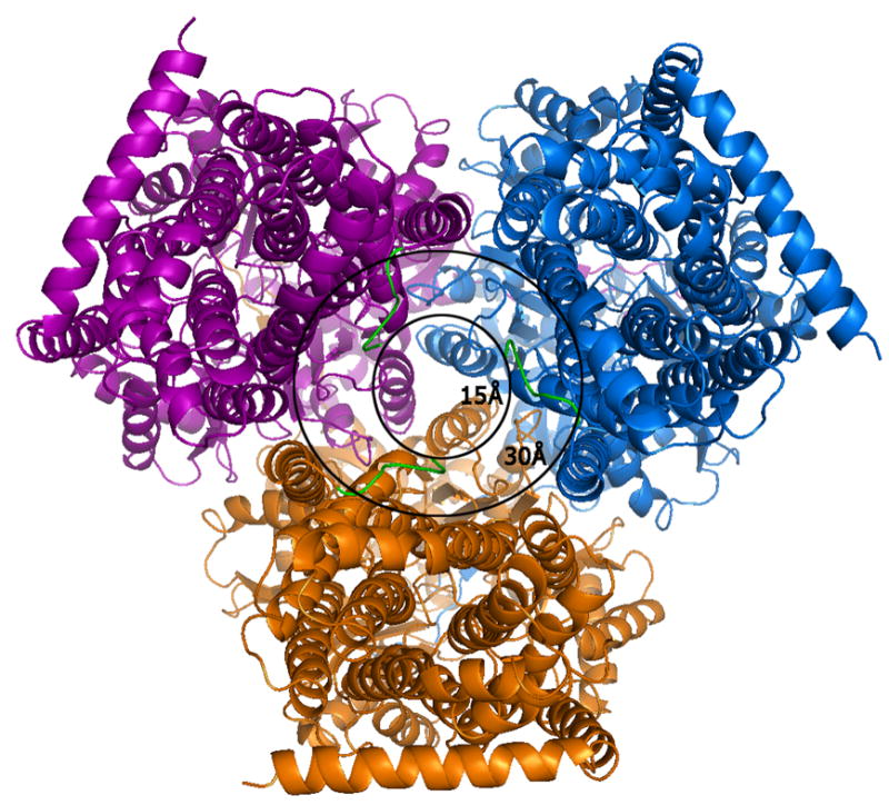
Figure 2.

A view of the AcrB trimer from the bottom, i.e.,the cytoplasmic face at a 90° view from that of Figure 1. The outer circle with a diameter of 30Å represents the transporter entry point size as reported in all previous crystal structures of AcrB in R32 space group lacking the first six amino acids. The extensions of the N-terminus (green) represent our model which completes the N-terminal model and reveals the actual cavity entry size of only ∼15Å. The smaller cavity opening and presence of conserved residues in the N-terminus implies a possible role of these initial residues in ligand capture and discrimination during cytosolic drug uptake.
