Figure 1.
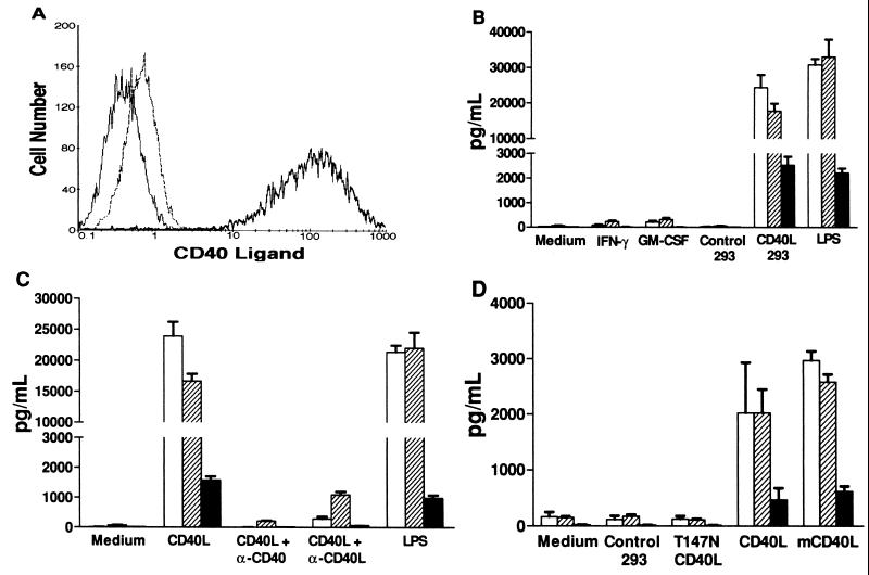
CD40L induction of β-chemokine production by macrophages. (A) Flow cytometry measurement of CD40L surface expression. Isotype control staining of CD40L-293 cells with PE-conjugated anti-CD8 control mAb (thin solid line). Expression of human CD40L on control 293 cells (thin broken line) and CD40L-293 cells (thick solid line) assessed by using PE-conjugated anti-CD40L mAb 24–31. (B) β-chemokine production by macrophages. MDM were cultured in medium alone, with added IFN-γ or GM-CSF, or with added control 293 cells or CD40L-293 cells. LPS was used as a positive control. The mean chemokine concentrations (picogram per milliliter) of the supernatants from quadruplicate wells 24 hr later are shown (±SD). □, MIP-1α; ▨, MIP-1β; ▪, RANTES. (C) Abrogation of CD40L stimulation by anti-CD40 or anti-CD40L mAbs. In an experiment similar to B, neutralizing mAbs were added at the initiation of the CD40L-293 cell-MDM cocultures. Anti-CD40 mAb blocked >99% and anti-CD40L mAb blocked 94% of the β-chemokine release. (D) Stimulation of macrophages by CD40L-bearing plasma membranes. Acellular preparations of membranes from control 293 cells, 293 cells expressing a nonfunctional mutant of human CD40L (T147N), human CD40L-293 cells (CD40L), and murine CD40L-293 cells (mCD40L) were added to MDM in an experiment similar to B.

