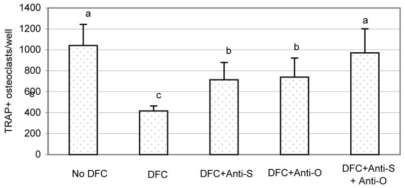
Fig. 5.

The bone marrow cells were co-cultured with the dental follicle cells in the presence of CSF-1 and RANKL, as well as anti-SFRP-1 (Anti-S) and anti-OPG (Anti-O) for 7 days. The bone marrow cell culture was then stained for TRAP+ osteoclasts, and TRAP+ osteoclasts per well were counted. Note that the presence of the dental follicle cells inhibited osteoclastogenesis but addition of either anti-SFRP-1 or anti-OPG in the culture increased osteoclast numbers. Addition of both antibodies essentially negated the inhibitory effect of the dental follicle cells. The bars with different letters between them indicate a statistically significant difference.
