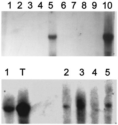
Figure 1.
Northern blot analysis of SCP-1 expression. Expression in normal tissues and tumor samples was tested. No expression is detectable in normal tissues (Upper) except for a high level expression in testis. Lanes: 1, brain; 2, PBMCs; 3, mitogen-stimulated PBMCs; 4, kidney; 5, testis; 6, spleen; 7, lung; 8, skeletal muscle; 9, liver; 10, testis. All lanes except lane 5 (5 μg) were loaded with 10 μg of total RNA. (Lower) Selected tumor tissues, proven to be positive for SCP-1 by specific RT-PCR, were retested by Northern blotting using 10 μg of total RNA, demonstrating significant transcripts levels, which were lower than the abundant transcript amounts detected in testis (lane T). Lanes: 1, breast cancer; 2, gastric cancer; 3, ovarian cancer; 4 and 5, renal cell carcinoma.

